


.bmp)


.bmp)
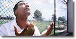
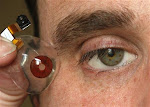
 In a fourth-floor lab at Harvard University, Adam Feinberg(Picture left) is peering through a low-magnification microscope and using a scalpel to cut out triangles and rectangles from a thin polymer.What's impossible to see with the naked eye is a one-cell-thick layer of heart tissue coating each shape. When Feinberg connects the petri dish holding the triangles and rectangles to a pacemaker, the tissue begins to rhythmically contract, and the shapes come alive-twisting, pinching, and even swimming through a solution.The pieces of "muscular thin films" are just a few millimeters long and only 30 micrometers thick; at first glance they resemble small worms you might find wiggling in a mud puddle. Kevin Kit Parker,(Picture right) the professor of biomedical engineering who heads the Harvard lab, jokes that he's planning to retire to the South, where he was raised, and sell them as customizable lures in a bait shop.But the experiment has entirely serious implications. Eventually, the patches of twitching tissue could be used as actuators for tiny robotic devices implanted in the body. The muscle cells would be fueled by sugar in the bloodstream and maintained by the same repair mechanisms that keep the heart pumping. Parker says the muscle-coated film could also be used to regenerate tissue damaged in heart attacks. But such applications are quite a way off, he says. In the nearer term, the devices could be used to help researchers monitor how experimental medicines change the behavior of heart muscle.Printing TissueThis is not the first time researchers have grown beating heart muscle in a dish. But Parker and Feinberg, a postdoctoral researcher in Parker's lab, have found ways to make the tissues far more powerful, contracting with the same strength as natural heart tissue.
The manufacture of the devices begins with a biological printing technique, developed by Harvard chemists, that can deposit proteins in microscopic patterns on various surfaces.Parker and Feinberg use the method to precisely organize the heart cells into working tissue.The process looks unremarkable. Working in a sterile laboratory hood, Feinberg arranges a few chunks of clear silicone rubber in a petri dish. The chunks are stamps patterned with an array of microscopic lines. The pattern was created by molding the stamps to a wafer of silicon etched using the same techniques that produce microchips. Onto each stamp Feinberg squirts a clear "ink" that contains a common protein called fibronectin. As the stamp dries, a thin layer of the protein forms. Holding a stamp with a pair of forceps, Feinberg presses it onto a round, silicone-coated glass coverslip, transferring proteins from the raised portion of the microscopic pattern to the silicone film.With the protein patterns stamped out and ready, Feinberg immerses the coverslip in a solution of young, still developing heart cells harvested from rats. The cells begin to adhere to the fibronectin, forming orderly lines. Feinberg then puts the cells and the protein-patterned coverslip, still immersed in the solution, into a body-temperature incubator. Over the next few days, the lines of fibronectin guide the cells' organization and further development. Long, fiberlike contractile units begin to form, guided by the cells so that they line up parallel to the lines of protein. If they weren't aligned this way, the cells would fight against each other as they contract rather than pulling in the same direction. The aligned cells, however, all contract along the same axis, much the way they do in natural heart tissue.
To read more go to:
As in the days of Noah....